Bifascicular Blocks
1.5% of patients may have a bifascicular block on the electrocardiogram.
This alteration is a combination of intraventricular conduction disturbances, which can carry a risk of progression to complete atrioventricular block, especially in patients with syncopal symptoms 1.
The 2009 AHA/ACCF/HRS Recommendations for the Standardization and Interpretation of the Electrocardiogram 1 do not recommend the term «bifascicular block» because of the great variation in anatomy and pathology producing such pattern. The committee recommends that each conduction defect be described separately in terms of the structures involved 1.
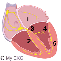
Anatomy of Bifascicular Block
Intraventricular Conduction
- 1- AV node and bundle of His
- 2- Right branch of bundle of His
- 3- Left branch of bundle of His
- 4- Anterior fascicle
- 5- Posterior fascicle
The ventricular conduction system can be classified as a three fascicles system. Consisting of the right bundle branch, and the anterior and posterior fascicles of the left bundle branch.
A bifascicular block is the combination of a blockade of two of these branches.
Types of Bifascicular Blocks
- Right bundle branch block + left anterior fascicular block.
- Right bundle branch block + left posterior ascicular block.
- Complete left bundle branch block: Equivalent to the block of both fascicles.
So, the bifascicular block has on the electrocardiogram the characteristics of right bundle branch block combined with the corresponding left fascicular block.
Remember: Left fascicular blocks are also called left hemiblocks
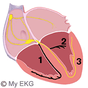
Right Bundle Branch Block + Left Anterior Fascicular Block
The most common of bifascicular blocks. The right bundle branch and the left anterior fascicle are blocked, so the depolarization of the ventricles is performed from the posterior fascicle of the left branch.
Right Bundle Branch Block + Left Anterior Hemiblock
- 1. Blocked right bundle branch.
- 2. Blocked left anterior fascicle.
- 3. Normal left posterior fascicle.
Characteristics of an Electrocardiogram with Right Bundle Branch Block and Anterior Hemiblock:
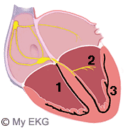
Right Bundle Branch Block + Left Posterior Hemiblock
The right bundle branch and the left posterior fascicle are blocked, so the depolarization of the ventricles is performed from the anterior fascicle of the left branch.
Right Bundle Branch Block + Left Posterior Hemiblock
- 1. Blocked right bundle branch.
- 2. Normal left anterior fascicle.
- 3. Blocked left posterior fascicle.
Characteristics of an Electrocardiogram with Right Bundle Branch Block and Posterior Hemiblock:
Bifascicular Blocks and complete AV block
In patients with bifascicular block in the electrocardiogram and episodes of syncope, there is the possibility that symptoms are secondary to an unknown advance atrioventricular block.
A more complex cardiology study should be performed in these patients, even, in some cases, it is advisable to perform an electrophysiological study to estimate the Hiss-ventricular interval (HV).
In patients with bifascicular block in the EKG without symptoms, the risk of advance atrioventricular block is low, but is recommended clinical and electrocardiographic monitoring.
Treatment of Bifascicular Block
Bifascicular blocks not require treatment except if the patient had syncope or an advance AV block is detected.
Permanent pacemaker implantation is indicated for complete AV block, or type II second-degree AV block. Also permanent pacemaker implantation is reasonable for syncope when other likely causes have been excluded 3.
previous | next
References
- 1. Surawicz B, Deal BJ et al. AHA/ACCF/HRS Recommendations for the Standardization and Interpretation of the Electrocardiogram Part III: Intraventricular Conduction Disturbances. Circulation. 2009; 119: e235-e240. doi: 10.1161/CIRCULATIONAHA.108.191095.
- 2. Epstein AE, DiMarco JP, Ellenbogen KA. ACC/AHA/HRS 2008 Guidelines for Device-Based Therapy of Cardiac Rhythm Abnormalities. A Report of the American College of Cardiology/American Heart Association Task Force on Practice Guidelines. Circulation. 2008; 117: e350-e408. doi: 10.1161/CIRCUALTIONAHA.108.189741.
If you Like it... Share it.