Left Fascicular Blocks
Left fascicular blocks, also called left hemiblocks, are conduction disturbances in one of two fascicles of the left bundle branch.
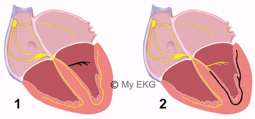
The left bundle branch is divided into two fascicles, the anterior fascicle and the posterior fascicle. Anterior fascicle transmits the electrical impulse to the upper and anterior regions of the left ventricle, the posterior fascicle to the inferoposterior regions of the left ventricle.
Left Fascicular Blocks (Left Hemiblocks)
- 1. Left anterior fascicular block
- 2. Left posterior fascicular block
In contrast to left bundle branch block, in left fascicular blocks, interruption of the stimulus occurs in only one of the fascicles.
Another difference with complete bundle branch blocks is, hemiblocks do not cause widening of the QRS complex (QRS complexes are narrow).
The main change of left hemiblocks in the electrocardiogram is a marked deviation of QRS axis.
Left anterior fascicular block has a marked left axis eviation and left posterior fascicular block has a marked right axis deviation (see how to determine the heart axis).
Left hemiblocks diagnosis is done in limb leads (difference with bundle branch blocks).
Left Anterior Fascicular Block (Left Anterior Hemiblock)
The main characteristic of the left anterior fascicular block in the electrocardiogram is a marked left axis deviation (-45º or more), without widening of QRS complex.
QRS complex has qR pattern in leads aVL.
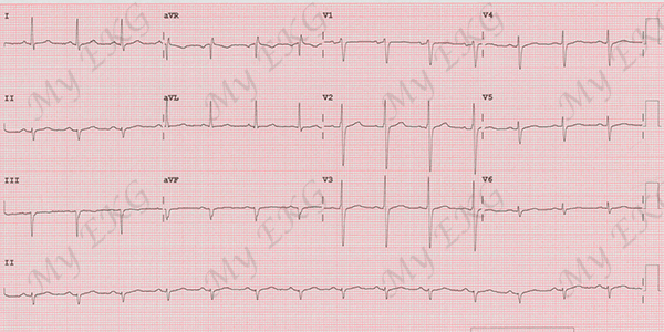
Left anterior fascicular block:
Sinus rhythm, 90 bpm, narrow QRS complex, left axis deviation, qR pattern in lead aVL.
Electrocardiogram of Left Anterior Fascicular Block
From AHA/ACCF/HRS Recommendations for the Standardization and Interpretation of the Electrocardiogram Part III: Intraventricular Conduction Disturbances 1.
- Frontal plane axis between −45° and −90°.
- qR pattern in lead aVL.
- R-peak time in lead aVL of 45 ms or more.
- QRS complex duration less than 120 ms.
These criteria do not apply to patients with congenital heart disease in whom left-axis deviation is present in infancy.
Explanation:
In anterior hemiblock, there is a delay in the activation of the anterior region of the left ventricle, which produces an initial, small wave. This wave is opposite to the direction of the blocked area, causing small r waves in inferior leads and small q waves in lateral leads.
Subsequently, the blocked area is depolarized, creating a big wave in that direction, causing deep S waves in inferior leads and tall R waves in lateral leads.
Related article: Anterior fascicular ventricular tachycardia.
Left Posterior Fascicular Block (Left Posterior Hemiblock)
The main characteristic of the left posterior fascicular block in the electrocardiogram is a right axis deviation (90º or more), without widening of QRS complex.
QRS complex has rS pattern in lateral leads and qR pattern in inferior leads.
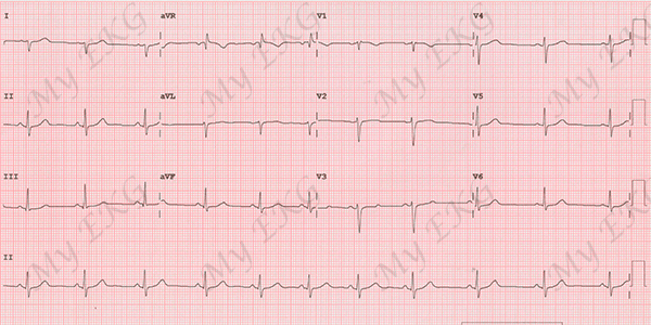
Left posterior fascicular block:
Sinus rhythm, 66 bpm, narrow QRS complex, right axis deviation, rS pattern in leads I and aVL, and qR pattern in leads III and aVF.
Electrocardiogram of Left Posterior Fascicular Block
From AHA/ACCF/HRS Recommendations for the Standardization and Interpretation of the Electrocardiogram Part III: Intraventricular Conduction Disturbances 1
- Frontal plane axis between 90° and 180° in adults.
- rS pattern in leads I and aVL.
- qR pattern in leads III and aVF.
- QRS complex duration less than 120 ms.
Explanation:
In posterior hemiblock, there is a delay in the activation of the posterior region of the left ventricle, which produces an initial, small wave. This wave is opposite to the direction of the blocked area, causing small r waves in lateral leads and small q waves in inferior leads.
Subsequently, the blocked area is depolarized, creating a big wave in that direction, causing deep S waves in lateral leads and tall R waves in inferior leads.
Unlike anterior hemiblock, the diagnosis of posterior fascicular block should only be done when other causes of right-axis deviation have been ruled out, as right ventricular overload or pulmonary embolism.
Related article: Posterior fascicular ventricular tachycardia.
Combination of Fascicular Blocks with Other Conditions
Left hemiblocks can be combined with other disturbances of the ventricular conduction system.
Combination of the Two Fascicular Blocks
Related article: Left bundle branch block.
The combination of the two left fascicular blocks causes a complete left bundle branch block.
If both fascicules of the left bundle branch are blocked, the ventricular depolarization is performed by the right branch, causing a widening of the QRS and characteristic changes in precordial leads.
More information: Left bundle branch block.
Fascicular Block with Right Bundle Branch Block
A left fascicular Block accompanied by a complete right bundle branch block on an electrocardiogram is called bifascicular block.
In bifascicular blocks they are affected two of the three branches of the intraventricular conduction system, so these patients have an increased risk of presenting high grade atrioventricular blocks, and in certain cases may require implantation of an electronic pacemaker.
previous | next
References
- 1. Surawicz B, Deal BJ et al. AHA/ACCF/HRS Recommendations for the Standardization and Interpretation of the Electrocardiogram Part III: Intraventricular Conduction Disturbances. Journal of the American College of Cardiology Mar 2009, 53 (11) 976-981. doi: 10.1016/j.jacc.2008.12.013.
If you Like it... Share it.


