Read and inform an electrocardiogram of a patient with a pacemaker is much more complex than write: "pacemaker rhythm".
As in a standard electrocardiogram (read how to read and electrocardiogram), the first thing to observe is the baseline heart rhythm.
Ignore for a moment the pacing spikes and concentrate on the baseline rhythm, in how the heartbeat is generated.
Search for P Waves
Native atrial activity:
- If native P waves are observed, it should be described as sinus atrial stimulation (or ectopic atrial stimulation).
P waves preceded by pacing spike:
- When P waves are preceded by pacemaker spikes, it should be described as atrial pacing.
In the absence of P waves:
- In the absence of P waves, it should be described as atrial flutter if "saw-tooth" waves are present, or atrial fibrillation if there are no atrial activity.
If you have understood this, you have mastered mastered the most difficult of reading an electrocardiogram with pacemaker.
The following step is to determine how the ventricles are stimulated. To do this, we should look at the QRS complexes.
Ventricular Stimulation
- Narrow QRS complexes without spikes: It should be described as native ventricular stimulation, and the description of the native QRS complex.
- Wide QRS complexes preceded by pacing spikes: It should be described as ventricular pacing with left bundle branch block pattern (QS in lead V1), right bundle branch block pattern (RSR' in lead V1), or QS pattern in all precordial leads.
The descriptions of initial rhythm and ventricular pacing must now be joined. For example:
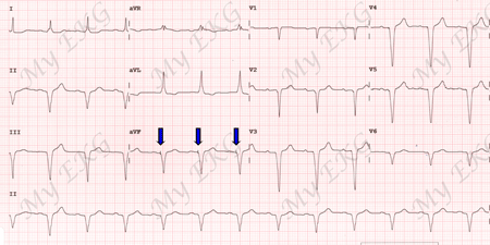
Sinus rhythm and ventricular pacing (blue) with left bundle branch block pattern.
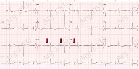
Atrial pacing (red) and native ventricular stimulation with normal PR interval.
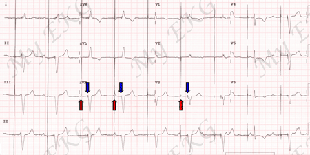
Atrial (red) and ventricular pacing (blue) with right bundle branch block pattern.
Related Article: Pacemaker malfunction on the EKG.


