Bundle Branch Blocks
Bundle branch blocks are disorders of electrical conduction distal to the bifurcation of the His bundle, causing changes in the depolarization of the ventricles.
This disturbance in ventricular depolarization causes marked changes in the QRS complex. In severe cases (complete bundle branch blocks), it causes an increase in the duration and changes in the morphology of the QRS complex.
The depolarization disorder also causes an alteration in ventricular repolarization. So the T wave also presents morphological changes in complete bundle branch block 1.
Anatomy of the Bundle Branch Blocks
Intraventricular Conduction
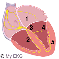
- 1- Atrioventricular ode and bundle of His.
- 2- Right bundle.
- 3- Left bundle.
- 4- Anterior fascicle.
- 5- Posterior fascicle.
The bundle of His divides into two branches:
- Right branch stimulates the right ventricle and the right third of the interventricular septum.
- The left branch stimulates the left ventricle and the left two-thirds of the interventricular septum.
While the right arm remains undivided, the left branch subdivides into two smaller branches ot fascicles.
- Anterior fascicle, which transmits the impulse to the anterior-superior region of the left ventricle.
- Posterior fascicle, which transmits the impulse to the posterior-inferior region of the left ventricle.
.
Any disturbance in one of these three branches causes conduction disorder and electrocardiographic abnormalities.
Complete Bundle Branch Blocks
A complete bundle branch block occurs when there is a complete obstruction of the impulses in the right branch or left branch before the subdivision.
On the electrocardiogram, the main characteristic of a complete bundle branch block is the QRS widening (greater than 0.12 s).
This is caused because the electrical stimulation depolarizes first the ventricle with normal branch, and then, through myocardium, depolarizes the ventricle with affected branch.
This increases the time of ventricular depolarization and therefore a widening of the QRS complex.
More information: Right bundle branch block, left bundle branch block.
Left Fascicular Blocks or Hemiblocks
The fascicular blocks occur when the interruption of the impulse is in one of the fascicles of the left bundle branch.
These blocks do not increase the time of ventricular depolarization. So the QRS complex is narrow.
Its main feature in the electrocardiogram is an important axis deviation.
More information: Left fascicular blocks.
Incomplete Bundle Branch Blocks
In incomplete bundle branch blocks, there is no interruption of the stimulus. The impulse is conducted by both branches, but more slowly through the affected branch.
On the electrocardiogram, they are observed with minimal widening of the QRS (between 0.1 s and 0.12 s) and morphological changes similar to bomplete bundle branch blocks.
Incomplete Right Bundle Branch Block:
Incomplete right bundle branch block is defined by QRS duration between 110 and 120 ms in adults. Other criteria are the same as for complete RBBB.
- QRS complex duration between 0.11 s and 0.12 s.
- rsr', rsR', or rSR' pattern in leads V1 or V2.
- S wave of greater duration than R wave in leads I and V6 in adults.
More information: Incomplete right bundle branch block.
Incomplete Left Bundle Branch Block:
- QRS complex duration between 110 and 120 ms in adults.
- Presence of left ventricular hypertrophy pattern.
- R peak time greater than 60 ms in left precodial leads.
- Absence of Q wave in leads I, V5, and V6 1.
More information: Incomplete left bundle branch block.
In both Incomplete bundle branch blocks could be ST segment or T wave abnormalities.
Disturbance of the Intraventricular Conduction
References
- 1. Surawicz B, Deal BJ et al. AHA/ACCF/HRS Recommendations for the Standardization and Interpretation of the Electrocardiogram Part III: Intraventricular Conduction Disturbances. Journal of the American College of Cardiology Mar 2009, 53(11): 976-981. doi: 10.1016/j.jacc.2008.12.013.
Next
If you Like it... Share it.


