Normal ST-Segment
The ST-segment, in normal conditions, is flat or isoelectric, although small variations up to 0.5 mm may be present.
To determine ST shift, the segment between previous T wave and the actual P wave (previous -TP segment) is used as reference. In cases where the T-P segment is not isoelectric the PR segment is used (check out the differences between intervals and segments).
Normal ST-segment elevation:
A mild ST-segment elevation, slight convex, with normal morphology, in right precordial leads, can be observed in healthy individuals. An ST convex elevation between 1 and 3 mm may also be present in cases of vagotonia and early repolarization, especially in precordial leads.
Normal ST-segment depression:
It is often observed during physical exertion, usually presenting a quick upslope crossing the isoelectric line.
Myocardial Ischemia: ST-Segment Abnormalities
The most important cause of ST-segment abnormalities (elevation or depression) is myocardial ischemia.
An electrical disturbance is originated in the myocardial tissue when a heart region is suffering from an important persistent ischemia, causing the ST-segment to shift on the EKG, either a upslope or a downslope, depending on the degree of coronary artery occlusion.
ST-Segment Elevation and Myocardial Ischemia
Acute ST-segment elevation on the electrocardiogram is one of the earliest signs of acute myocardial infarction, generally related to acute and complete coronary artery occlusion.
To diagnose ST-segment elevation myocardial infarction (STEMI) this elevation must be persistent in at least two adjacent leads.
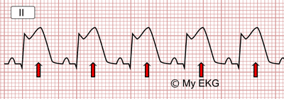
ST-segment elevation in the electrocardiogram
ST-Segment Depression and Myocardial Ischemia
Acute ST-segment depression is, as elevation, a sign of myocardial injury. It generally correlates with incomplete coronary artery occlusion (see NSTE-ACS). As with elevation, ST-segment depression must be present in at least two adjacent leads.
It could be persistent or transient, and it is a sign of disturbances during ergometry. It also appears as a reciprocal image in leads no affected by a STEMI.
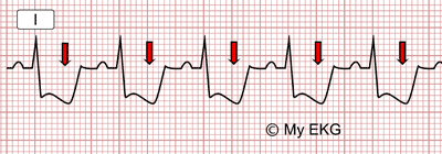
ST-segment depression on the Electrocardiogram
Reciprocal ST Segment Depression
During a STEMI, leads with ST elevation and leads ST depression may appear on the same EKG. This is called reciprocal image.
Leads with ST depression are leads which are not affected by coronary occlusion, they just mirror ST segment elevation, hence the name reciprocal image.
It is important to note that on an EKG that presents both ST elevation and depression, ST elevation leads are the ones reflecting myocardial injury, thus determining location and extent of infarction.
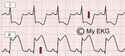
Reciprocal changes:
ST elevation in inferior leads (DII) and ST depression in lateral leads (DI)


