Steps to Recognize Normal Sinus Rhythm
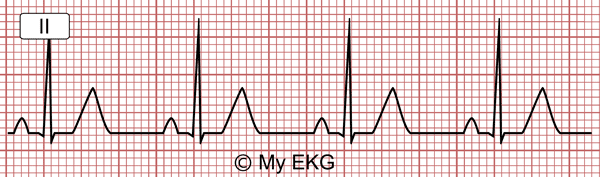
Sinus rhythm at 75 bpm.
Heart Rate Between 60 bpm and 100 bpm
Sinus rhythm has a heart rate higher than 60 bpm and lower than 100 bpm. If heart rate is lower than 60 bmp it is called sinus bradycardia, if heart rate is higher than 100 bpm it is called sinus tachycardia.
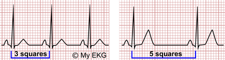
Sinus rhythm: Minimum R-R interval (100 bpm = 3 squares) and maximum R-R interval (60 bpm = 5 squares).
As sinus rhythm is regular (read next step) if the distance between two QRS complexes (R-R interval) complex is larger than 3 big squares and less than 5 big squares, the heart rate is normal.
Sinus Rhythm is Regular
In sinus rhythm, the RR intervals are constant, or almost constant, but, how to determine if the rhythm is regular?
Sometimes, it is easy to determine if the rhythm is regular, but in cases of doubt we can measure with a ruler or a compass.
A simple tip is to mark 3 QRS complexes on a blank sheet of paper and move the marks to the next QRS complexes, if they match, the R-R interval is constant and the rhythm is regular.
Minor variation in the P-P intervals is present in most subjects. When this variability is more accentuated, the term sinus arrhythmia is used.1
Sinus P Waves
In sinus rhythm atrial activation begins in the sinus node. The sinus node is located in the upper-right corner of the heart. The atrial depolarization spreads from right to left and downward toward the atrioventricular node.
Therefore, the stimulus move away from the lead aVR and is directed toward the lead II (P wave axis is directed toward lead II).
The sinus P wave is positive in all leads except aVR where it is negative, and V1, where it is biphasic.
If P wave do not comply this characteristc, the patient could have an atrial ectopic rhythm (the stimulus arise from a different location) or a misplacement of the limb lead electrodes.
A P Wave for Each QRS Complex
In sinus rhythm each P wave is followed by a QRS complex.
If some of the P waves are not followed by a QRS complex (not conducted P waves) would likely be a second or third degree AV block, therefore, this rhythm is not a normal sinus rhythm.
PR Interval higher than or equal to 0.12 s
Short PR intervals with delta waves are signs of the existence of an accessory pathway (read Wolff-Parkinson-White).
It is not considered normal sinus Rhythm because the stimulus is conducted by a structure outsider the normal conduction system.
There may also be short PR interval in low atrial rhythm or in retrograde atrial conduction due to a junctional rhythm, but neither is sinus rhythm.
Normal Sinus Rhythm and Long PR interval:
There is no general agreement on whether the long PR interval (>0.2 s) rules out normal sinus rhythm.
The majority of authors accept it as Normal Sinus Rhythm, because the stimulus is conducted by normal conduction system.
It may be described as sinus rhythm with first degree AV block or long PR interval.
Summary
The normal sinus rhythm has these electrocardiographic characteristics:
- Heart rate between 60 and 100 bpm (R-R interval between 3 and 5 big squares)
- RR interval must be constant (similar R-R intervals).
- Positive P wave in lead II and negative in lead aVR.
- Each P wave is followed by a QRS complex
- PR interval must be ≥0.12 seconds.
Sinus Rhythm with Other Disturbances
When we speak about normal sinus rhythm, the adjective “normal” refers only to heart rhythm, it does not mean that the complete EKG is normal.
For example, normal sinus rhythm can be described in the presence of bundle branch block or signs of acute myocardial infarct.
It is a different matter when an initial sinus rhythm is associated with other heart rhythm disturbance as AV blocks, conduction via accessory pathway or pacemaker pacing.
In these conditions, the diagnosis of sinus rhythm should be restricted to atrial rhythm only.
In these cases is possible to determine sinus rhythm because P waves fulfill the first three criteria described above, replacing R-R interval with P-P interval.
- Heart rate between 60 and 100 bpm (P-P interval between 3 and 5 big squares).
- P-P interval must be constant (similar P-P intervals).
- Positive P wave in lead II and negative in lead aVR.
Sinus Rhythm with 2nd or 3rd degree AV Blocks
Atria are depolarizated by sinus node, but some (2nd degree AV block) or all (complete AV block) stimuli are not conducted to the ventricles.
In 2nd degree AV block, not all sinus P waves are followed by QRS complexes because there is an intermittent failure of the AV conduction.
In complete AV block, sinus P waves are dissociated from QRS complexes. No P waves are conducted to the ventricles because there is a complete block of the AV conduction.
It may be described as sinus rhythm with second or third degree AV block.
Sinus Rhythm with Ventricular Pacemaker Pacing
The patient with electrical pacemaker may have sinus rhythm in the atria with ventricular pacing.
In dual-chamber pacemakers programmed in DDD mode ventricular pacing must follow the sinus P wave.
In single-chamber pacemaker (usually in VVI mode), if there were sinus activity, ventricular pacing would be independent of atrial rhythm.
In both cases we may describe the EKG as sinus rhythm with ventricular pacing by electrical pacemaker.
In patients with pacemaker, if pacing is inhibited by sensed spontaneous cardiac depolarizations, we can describe normal sinus rhythm.
Sinus Rhythm with Pre-excitation Syndrome
Patients with accessory pathway have a double atrioventricular conduction: AV node and accessory pathway, this cause a short PR interval and a delta wave on the EKG (read pre-excitation syndromes).
It is not a normal sinus rhythm if signs of pre-excitation are seen on the electrocardiogram.
It may be described as sinus rhythm with short PR interval with delta wave.
Sinus Rhythm with Ventricular Arrhythmias
During a ventricular tachycardia or an accelerated idioventricular rhythm, a basal sinus rhythm can be seen with atrioventricular dissociation. P waves are usually difficult to distinguish..
The P wave rate is lower than ventricular rate.
The A-V dissociation in a broad QRS complex tachycardia is one of the diagnostic criteria of ventricular tachycardia.


