Pacemaker Malfunctions on the Electrocardiogram
The electrical pacemaker, as every device, can present alterations in its operation. In these cases the electrocardiogram is the first tool with which we have to detect them.
The dysfunctions of pacemakers can be classified in disorders of the stimulation (generation or transmission of the stimulus), or the sensing (absence or bad detection of cardiac stimulation).
Problems with Pacing
Pacing disorders occur when the electrical stimulation of the device does not occur or is not transmitted to the myocardium.
The main causes are often the fracture or displacement of the lead, battery depletion, electrolyte disturbance or antiarrhythmic treatment (in these last two the function of the pacemaker is correct but is not able to stimulate the muscle).
Remember: The full or partial absence of spikes does not mean a problem of pacing, it can be inhibited by a patient's heart rhythm with a higher heart rate.
Absence of Pacing Spikes
The electrocardiogram shows a partial or total absence of pacemaker spikes despite the existence of conduction problems.
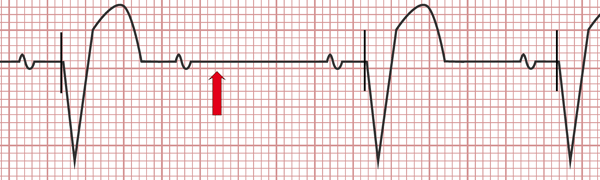
Electrocardiogram of pacemaker malfunction: Pacing spikes are absent (red arrow).
Conduction disorders (AV blocks, sinus pauses) without pacing are observed on the electrocardiogram.
Capture Failure
On the surface EKG, pacing spikes are present, but they are not followed by a cardiac electrical activity (P waves or QRS complexes).
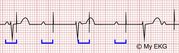
Complete AV block with a DDD pacemaker malfunction due to capture failure:
The device senses P waves (blue) without ventricular capture.
On the electrocardiogram, the baseline rhythm of the patient is observed along with pacing spikes which do not produce P waves or QRS complexes.
Extracardiac Stimulation:
It usually occurs when there is a lead dislodgment it is out of the heart stimulating other muscle groups (skeletal muscles or diaphragm).
On the electrocardiogram is usually seen absence of pacing spikes. To confirm the diagnosis, it is enough with a chest x-ray that allows you to see the lead displaced.
Problem with Sensing
In disorders of sensing, the pacemaker does not correctly recognize cardiac stimuli, either by excess or by default.
Improper programming, alterations of the circuit, displacement or fracture of the lead, are the main causes of sensing disorders.
Oversensing Problems
The pacing is inhibited (do not paces) because the pacemaker senses cardiac signals (wrongly detects T or P waves as QRS complexes) or extracardiac signals (artifacts, myopotential).
On the EKG, pacing spikes are absent, or the pacing rate is lower than the programming for the device.
Undersensing Problems
The pacemaker fails to detect spontaneous myocardial depolarization, ant it paces as if there were no underlying rhythm.
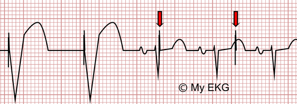
Undersensing:
Pacing is not inhibited by the QRS complexes and produces spikes (red arrows).
On the electrocardiogram, there are pacing spikes with a regular rhythm. These spikes may or may not be followed by a QRS complex, and there may be fusion beats.
Pacemaker-mediated Tachycardia
Endless-loop Tachycardia
It is a re-entrant tachycardia, which occurs in the dual-chamber pacemakers.
The ventricular stimulus is conducted by the normal condution system (retrograde conduction ) and it is sensed as native atrial activity with subsequent ventricular pacing; this ventricular stimulus returns to the atria by the normal condution system, closing the circuit.
The endless-loop tachycardia is observed on the electrocardiogram as a tachycardia with pacemaker pacing close to the maximum rate limited by the pacemaker programming.
Placing a magnet on the device results in the termination of tachycardia by suspending the atrial sensing.
Tracking an Atrial Tachyarrhythmia by Dual Chamber Pacemaker
Onset of atrial tachycardia (atrial flutter, atrial fibrillation or atrial tachycardia) that is tracked by the pacemaker.
On the EKG, it is observed as a tachycardia with ventricular pacing, in the case of atrial fibrillation can be seen irregular ventricular pacing.
Sensor Induced Tachycardia
Sensor induced at tachycardia occurs when limb movement during exercise, rapid hyperventilation, vibrations, loud noises, fever, acidosis, or electrocautery during surgery causes the sensor of a modern pacemaker to fire at an inappropriately rapid rate.
On the electrocardiogram, the ventricular rate is at the upper limit of the pacemakers range at 160-180 bpm.
If you Like it... Share it.


