Left fascicular ventricular tachycardia is the most common of the idiopathic ventricular tachycardias originating in the left ventricle. It is approximately 10% of all idiopathic VTs (the most common idiopathic VT are right ventricle outflow tract tachycardia) 1.
Left fascicular ventricular tachycardias usually ocurr in healthy patients, mainly as sustained monomorphic VT, with right bundle branch block pattern on the EKG and with narrower QRS complexes than the rest of the monomorphic ventricular tachycardias (QRS complexes less than 140 ms).
They usually respond to treatment with verapamil, which is why they are also called verapamil-sensitive ventricular tachycardias.
These arrhythmias usually respond to treatment with verapamil, which is why they are also called verapamil-sensitive ventricular tachycardias.
Posterior Fascicular Ventricular Tachycardia
It is the most frequent form of left fascicular ventricular tachycardia (90%), in this VT the posterior fascicle is involved.
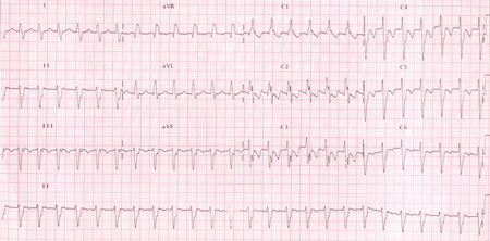
Image from Ramprakash B et al. Catheter Ablation of Fascicular Ventricular Tachycardia 2.
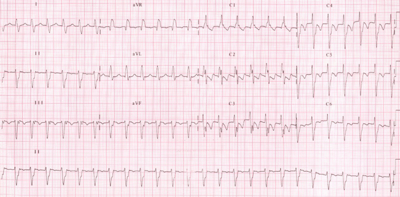
Posterior fascicular ventricular tachycardia:
Wide QRS complex tachycardia with right bundle branch block pattern and left-axis deviation, around -90º.
The electrocardiogram shows:
Anterior Fascicular Ventricular Tachycardia
It represents approximately 10% of left fascicular ventricular tachycardia, in this TV is involved the anterior fascicle.
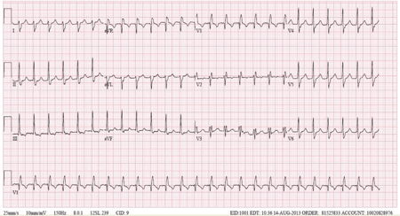
Image from Bennin C et al. Idiopathic Left Anterior Fascicular Tachycardia Presenting after Aortic Valve Replacement 3.
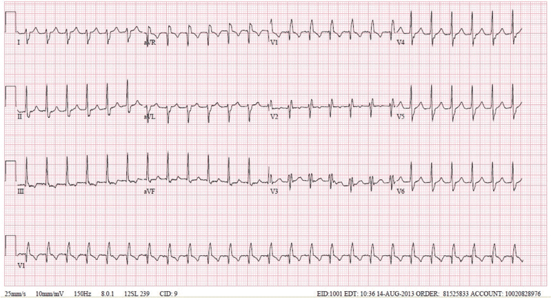
Anterior fascicular ventricular tachycardia:
Wide QRS complex tachycardia with right bundle branch block pattern and right-axis deviation, around 120º.
The electrocardiogram shows:
Upper Septal Fascicular Ventricular Tachycardia
It is an exceptional form of presentation, involving a third fascicle of the conduction system: the septal fascicle.
The electrocardiogram shows:
- Right bundle branch block pattern with QRS complexes less than 140 ms.
- Normal heart axis.
- It may also be observed as narrow QRS complex tachycardias, even similar to sinus QRS, or with incomplete bundle branch block pattern (right or left), with normal heart axis or with right-axis deviation.
Upper septal fascicular ventricular tachycardia may present as a narrow QRS ventricular tachycardia.
Treatment of Idiopathic Fascicular Left Ventricular Tachycardia
Acute Management
As in all arrhythmias, acute treatment depends on the hemodynamic stability of the patient.
In case of hemodynamic instability, the treatment of choice is electrical cardioversion.
In stable patients, first line treatment for left fascicular tachycardias is verapamil IV (bolus 10 mg given for over 1-2 minutes) 4 5.
Remember: fascicular left ventricular tachycardia are also called verapamil-sensitive tachycardias.
It is important to remember that the verapamil administration can cause hemodynamic disorders, so it should be administered only if the patient is stable and there is an established diagnosis.
Fascicular left ventricular tachycardias usually do not respond to beta-blockers, adenosine, or vagal maneuvers 5.
Long-term Management
Oral verapamil is a therapeutic option in patients with mild symptoms and rare episodes of fascicular tachycardia.
In patients with severe symptoms, frequent episodes or poor response to treatment with verapamil the first line treatmen is catheter ablation of the tachycardia 4 5.