Atrial Septal Defects (ASD) on the Electrocardiogram
Authors: Eduardo Consuegra Llapur, Ernesto C. Amalfi Aguilera.
Atrial septal defect (ASD), also known as interatrial communication, is the fifth congenital heart disease in order of frequency.
The International Society for Nomenclature of Paediatric and Congenital Heart Disease (ISNPCHD) has defined an interatrial communication as a congenital cardiac malformation in which there is a hole or pathway between the atrial chambers 1.
Atrial septal defect is classified according to the area of the septum in which it is located. In general, its anatomical forms are:
- Ostium secundum: they are the most common type of ASD, constituting 80% of the cases. It is located in the area of the fossa ovalis.
- Sinus venosus: they comprise 5% of ASD. They are locatedin the superior or inferior region of the interauricular septum, near the superior or inferior vena cava respectively.
- Ostium primum: they are the second most frequent ASD, up to 15%. It is located near the crux of the heart, and is usually associated with other defects such as mitral cleft, presence of common atrioventricular valve, or inlet ventricular septal defect.
Ostium Secundum and Sinus Venosus
Normally, these cardiopathies do not show signs of clinical relevance until the child is over 3 or 4 years old.
The electrocardiogram presents the same findings in the ostium secundum and venous sinus forms.
The left to right shunt that occurs at the atrial level causes volume overload of the right atrium and the right ventricle, as well as increased pulmonary flow; this is observed with high P waves in all leads of the EKG except aVR.
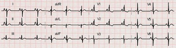
Ostium secundum ASD: high P waves, right bundle branch block, deep S waves in V6, and right axis deviation.
A right bundle branch block pattern is common (rSr' pattern in leads V1 and V2). Deep S waves in lead V6 are also common, representing a right ventricle dilation.
The electrocardiogram usually has a right axis deviation.
The Cabrera and Monroy overload criteria are clearly applied in these two forms of atrial septal defect. In these cases, diastolic overload of the right ventricle is expressed by a right bundle branch block pattern with negative T waves in leads V1 and V2.
Ostium Primum
The ostium primum is a defect in the atrial area of the atrioventricular septum. That is, the antero-inferior zone of the atrial septum.
It is currently classified among the atrioventricular septal defects, being this one of a partial type (partial atrioventricular canal, partial endocardial cushion defect).
This defect does not have great differences in its clinical picture with the other ASDs, but it has electrocardiographic characteristics that may facilitate its diagnosis.
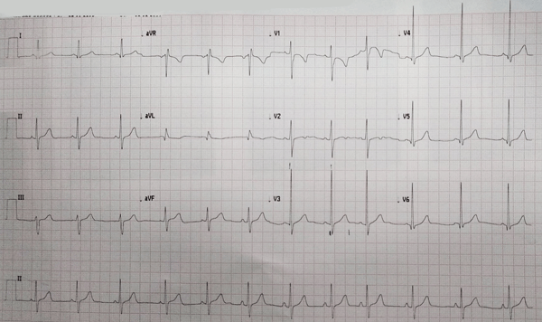
Ostium primum ASD: a 22-month-old patient EKG with electrical axis at -5° (left axis deviation for his age).
It is most often associated with cleft of the mitral septal leaflet, so it may be accompanied by varying degrees of mitral regurgitation.
This defect frequently causes displacement of the sonduction system, resulting in a left axis deviation.
Clinical suspicion of ASD with left axis deviation will allow us to make the diagnosis of this defect.
References
- 1. Franklin RC, Béland MJ et al. Nomenclature for congenital and paediatric cardiac disease: the International Paediatric and Congenital Cardiac Code (IPCCC) and the Eleventh Iteration of the International Classification of Diseases (ICD-11). Cardiol Young. 2017; 27: 1872-1938. doi: 10.1017/S1047951117002244.
- 2. Allen HD, Gutgesell HP, Clark EB, Dricoll DJ: Moss and Adam’s Heart Disease and Infants, Children and Adolescents Incluiding the Fetus and Young Adult, 6th ed. Philadelphia, PA, Lippincott Williams & Wilkins, 2001.
- 3. Rijnbeek PR, Witsenburg M, et al. New normal limits for the paediatric electrocardiogram. Eur Heart J. 2001 Apr; 22(8):702-11. doi: 10.1053/euhj.2000.2399.
- 4. Yavuz T, Nisli K, Oner N, Dindar A, Aydogan U, Omeroglu RE, Ertugrul T:The effects of surgical repair on P-wave dispersion in children with secundum atrial septal defect.Adv Ther. 2008 Aug;25(8):795-800.
If you Like it... Share it.


