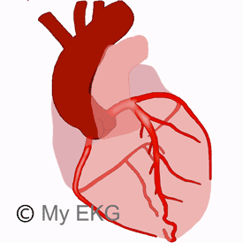
Coronary Arteries Anatomy
The coronary arteries are the arteries in charge of nourishing the entire heart. Their name comes from the Latin word coronarĭus, which means in the shape of a crown, due to the wat that they surround the heart.
There are two coronary arteries, the right coronary artery and the left coronary artery which emerge from the ascending aort, from the right and left aortic sinuses respectively.
The left main coronary artery has a short common trajectory, branching into the left anterior descending artery and the circumflex artery.
This is why the coronary system is usually considered a system with three major arteries, the left anterior descending artery, the circumflex artery and the right coronary artery. They are important in the diagnosis of ischemic heart disease.
Left Coronary Artery
Left Main Coronary Artery
The left main coronary artery is the beginning of the left coronary artery. It originates in the left posterior aortic sinus and passes behind the pulmonary artery. It divides into two branches: the left anterior descending artery and the circumflex artery.
The length of the left main coronary artery is highly variable, and can reach up to 20 mm. In some patients, it does not exist and the left anterior descending artery and circumflex artery originate independently in the left posterior aortic sinus.
Left Anterior Descending Artery (LAD)
Also called the anterior interventricular artery. It originates from the fork at the left main coronary artery, passes to the left of the pulmonary artery and runs through the anterior ventricular groove to the apex.
In most patients, it extends past it and ends in the distal third of the inferior wall (recurrent left anterior descending artery).
The LAD is the artery that irrigates the most territory of the left ventricle, the entire anterior wall and part of the lateral wall of the left ventricle are nourished from it, as well as the anterior two thirds of the septum, part of the right ventricular outflow tract and, in some patients, the apical segments of the inferior wall.
Several branches originate from the left anterior descending artery, the most important of which are the diagonal and septal branches.
Conus arteriosus artery:
Irrigates the right ventricular outflow tract (conus arteriosus), it usually communicates with its homonym coming from the right coronary artery.
Diagonal branches:
These are directed towards the lateral wall, which is irrigated along with the left marginal arteries coming from the circumflex artery.
Most frequently, there are one or two large diagonals present, but their number may vary.
Septal branches
Small branches that penetrate perpendicularly into the interventricular septum irrigating its two previous thirds. They may also vary greatly in number.
The exit order of the diagonal and septal arteries is different in each patient.
Circumflex Artery
It originates from the fork in the left main coronary artery and flows by the left coronary sulcus, bordering the heart towards its posterior region, in the direction of the posterior interventricular sulcus.
The posterior descending artery originates in the circumflex artery for 15% of patients, which is known as left-dominant; in the rest of the cases, the circumflex usually ends up near the posterior interventricular sulcus in a diffuse way.
The circumflex artery irrigates the lateral and posterolateral wall of the left ventricle, the lateral and posterior wall of the left atrium and, if there is left dominance, also irrigates the inferior wall of the left ventricle.
Along the way, the circumflex artery branches towards the left atrium and the lateral wall of the left ventricle.
Atrial branches:
They are small branches that irrigate the lateral and posterior walls of the left atrium.
Left marginal arteries:
Also called obtuse marginal arteries. They originate in the circumflex artery going towards the left margin of the heart (obtuse margin) irrigating the lateral and posterolateral wall of the left ventricle.
Posterior descending artery:
Also called posterior interventricular artery. It originates in the circumflex artery in 15% of the patients and runs through the posterior interventricular sulcus ending, in most cases, in the distal third of the inferior wall.
Ramus Intermedius
Up to one-third of patients have a trifurcated left main coronary artery. This gives the left anterior descending artery, the circumflex artery and a smaller artery exactly in between them which is called ramus intermedius.
This branch is directed towards the lateral wall of the heart, serving as a diagonal branch or a left marginal artery.
Right Coronary Artery
It originates from the right posterior aortic sinus and runs through the right coronary sulcus, surrounding the heart, until the posterior interventricular sulcus, where, in 85% of patients, it yields the posterior descending artery (right-dominant).
The right coronary artery branches off several times in its path. In most patients, it branches the sinoatrial node artery and then produces branches directed towards the lateral wall of the right ventricle (right marginal branches).
Conus arteriosus artery:
It irrigates the conus arteriosus or right ventricular outflow tract. It usually communicates with its homonym from the left anterior descending artery.
Sinoatrial nodal artery:
it originates from the right coronary artery in up to 60% of patients, in the rest it comes from the circumflex artery. It goes through the anterior wall of the right atrium to the superior vena cava ostium, surrounding it and ending at the sinus node.
Right marginal branches:
They originate from the right coronary artery in the direction of the lateral wall (acute margin) of the right ventricle.
Posterior descending artery:
In 85% de los pacientes it originates in the right coronary artery, passes through the posterior interventricular sulcus and ending, in most cases, before the distal third of the inferior wall. The artery of the AV node originates from it as well as several small septal branches that irrigate the posterior third of the interventricular septum.
AV node artery:
It usually arises from the right coronary artery and less frequently from the left circumflex artery, depending on which artery crosses the posterior interventricular sulcus.
Posterolateral branches:
These are not present in all patients. They are the continuation of the right coronary beyond the posterior interventricular sulcus. They irrigate the posterior wall of the left ventricle.
Coronary Artery Dominance
Coronary artery dominance is defined by the vessel which gives rise to the posterior descending artery.
Coronary circulation can be classified as right-dominant when the posterior descending artery originates from the right coronary artery (85% of patients), and left-dominant when it originates from the circumflex artery (15%).
Important: The artery that irrigates the most cardiac territory is the left coronary artery, even when there is right dominance..
Anatomic Variations of the Coronary Arteries
We have described the main arteries and their main branches, but we must remember that the anatomy of the coronary arteries varies from one individual to another.
Most frequent variations:
Posterior descending artery:
It indicates coronary dominance; it can come from the right coronary artery or the circumflex artery.
When the right coronary artery is dominant, it usually continues through the posterior interventricular sulcus (posterolateral branch), while the distal circumflex has a small diameter and does not reach the crux of the heart (union of the coronary sulcus with the interventricular sulcus).
On the contrary, when the circumflex artery is dominant, the right coronary artery is usually small and of little relevance, unless it presents some large right marginal branch.
Diagonal branches and left marginal arteries:
These arteries share a similar area of irrigation. So it is normal that if the diagonal branches are big, there are few left marginal arteries and these typically have small diameters.
Conversely, when the left marginal arteries are large, there are usually few diagonal branches and they tend to be have small diameters.
Recurrent left anterior descending artery:
In certain patients the left anterior descending artery borders the apex and continues through the posterior interventricular sulcus to its middle third, irrigating much of the lower wall.
In these cases, the posterior descending artery usually has a small diameter and is short.
Left anterior descending artery bifurcation:
Some patients present a bifurcation of the left anterior descending artery, presenting a large septal branch, from which all septal arteries originate, and a more lateral branch from which the diagonal branches originate.
Coronary Collateral Circulation
When a total or subtotal occlusion occurs in a coronary artery, the coronary flow distal to the occusion is interrupted.
When the occluded artery receives blood from another artery or from the same artery through a branch that has not been affected by the occlusion, it is called coronary collateral circulation.
This is produced through the communication of distal branches, usually of small diameter.
Coronary collateral circulation is of key importance in ST-segment elevation myocardial infarction, where its presence positively affects prognosis by reducing the degree of myocardial ischemia and alterations in myocardial contractility.
Collateral circulation is not always present and its location is highly variable.
If there is no coronary disease, it cannot be observed in the coronariography, but in the case of total or subtotal occlusion of an artery it is seen as part of this artery filled with contrast from its distal regions.
Collateral Circulation Between Different Arteries
Conus arteriosus arteries: Union of the distal segments of the Conus arteriosus arteries from the left anterior descendant and the right coronary arteries.
Septal branches: Communication between the septal branches of the left anterior descending artery and those of the posterior descending artery.
Diagonal branches – left marginal arteries: Union of small arteries between the diagonal branches and left marginal arteries on the lateral wall of the left ventricle.
Left anterior descending artery - posterior descending artery: Both arteries can communicate in the apical region.
Right marginal branches – left anterior descending artery: The distal segments of the right marginal branches can be joined to small branches of the left anterior descending artery.
Left marginal arteries – posterolateral branches: Usually share territory (posterolateral wall) so it is not uncommon for them to communicate with each other.
Collateral Circulation From the Same Artery
There may also be collateral circulation between the branches of the same artery, when a branch that originates before the occlustion site communicates with the distal segments of the same artery.
- Diagonals – left anterior descending artery.
- Left marginal arteries - circumflex artery.
- Right marginal branches – posterior descending artery or posterolateral branches (right coronary artery).
Collateral circulation allows the region affected by coronary occlusion to receive blood flow, which is sometimes sufficient to reduce the degree of ischemia.
It can even prevent the characteristic changes of coronary occlusion (see STEMI) and guarantee the perfusion of a large part of the myocardium until coronary reperfusion is performed.
That is why its presence is a factor that decreases the magnitude and complications of an acute infarction.
If you Like it... Share it..