Left Atrial Enlargement on the Electrocardiogram
The passage of the electrical stimulus through the atria is reflected in the electrocardiogram as the P wave.
The normal P wave measures less than 2.5 mm (0.25 mV) in height and less than 0.12 s in length (3 small squares).
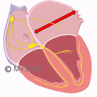
It is also composed of two components, an initial component where the depolarization of the right atrium is observed and a final component caused by the depolarization of the left atrium.
This difference is more striking in the lead V1 where the P wave has a biphasic morphology, with a first positive component (right atrium) and a second negative component (left atrium) 1.
Left Atrial Enlargement on the EKG
As it is to be supposed, the dilation of the left atrium produces, in most cases, changes in the P wave, especially in its final component.
As the left atrium depolarizes after the right atrium, an enlargement thereof will cause a longer duration of the depolarization time and therefore a widening of the P wave, greater than 0.12 s.
Sometimes the right and left component of the P wave are separated slightly giving the P wave a form of "letter m" lower case, classically called P mitrale.
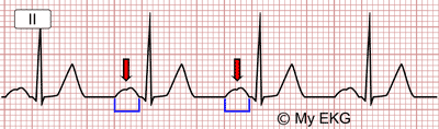
Left Atrial Enlargement:
Wide P wave, greater than 0.12 s, P mitrale (red arrow).
In addition, in lead V1, the depth of the negative final component is greater than the height of the initial part.
Summarizing: The most striking sign of the left atrial enlargement is a wide P wave, greater than 0.12 s or 3 small squares, with a predominance of the negative final component in lead V1.
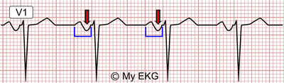
Left atrial enlargement:
Wide P wave with prominent negative component.
It is important to note that in patients with ischemic heart disease, wide P waves with a left atrium of normal dimensions can be observed, probably due to a delay of the atrial conduction.
The presence of electrocardiographic signs of left atrial enlargement is one of the criteria for the diagnosis of left ventricular hypertrophy (LVH), this is one of the few signs of LVH detectable on the EKG in patients with right bundle branch block (read left ventricular hypertrophy).
Causes of Left Atrial Enlargement
Aging itself causes left atrial growth, probably in relation to structural changes in the atrial tissue.
Obesity has also been related to left atrial enlargement, although the mechanism is not very clear 2.
left ventricular hypertrophy is clearly related to the left atrial enlargement, so those causes that cause LVH as hypertension, aortic stenosis or hypertrophic cardiomyopathy can lead to left atrial enlargement.
Alterations of the mitral valve are the classic causes of left atrial enlargement, both mitral stenosis due to increased pressure, and mitral insufficiency due to volume increase.
Aortic insufficiency generates left cavities overload propitiating left atrial and left ventricular enlargement.
Atrial fibrillation is both cause and effect of left atrial enlargement, although the presence of AF on the EKG makes it difficult to determine left atrial enlargement signs, because P waves are absent 4.
Causes of Left Atrial Enlargement
Left Atrial Enlargement and Interatrial Blocks
Related article: Bayés syndrome and interatrial blocks.
The interatrial block pattern presents a P wave widening that is frequently bimodal, which often leads to interpretation as left atrial enlargement, but these two electrocardiographic patterns are two different entities 5.
In these cases, it is the morphology of the P wave in lead V1 that allows us to determine if there is a left atrial enlargement associated with interatrial block.
The presence of a negative final component of the P wave in lead V1 greater than 40 ms may indicate left atrial enlargement 5.
In any case, the association between interatrial block and left atrial enlargement is relatively frequent. In fact, it has been considered that the bimodal P wave is better explained because of underlying interatrial block than the longer distance that the impulse has to go across 6.
More information: Bayés syndrome and interatrial blocks.
References
- 1. Surawicz B, Knilans TK. Chou’s electrocardiography in clinical practice, 6th ed. Philadelphia: Elservier; 2008.
- 2. Edhouse J, Thakur RK, Khalil JM. Conditions affecting the left side of the heart. BMJ 2002;324:1264. doi: 10.1136/bmj.324.7348.1264.
- 3. Vaziri SM, Larson MG, Lauer MS, et al. Influence of Blood Pressure on Left Atrial Size. The Framingham Heart Study. Hypertension. 1995; 25: 1155-1160. doi: 10.1161/01.hyp.25.6.1155.
- 4. Bombelli M, Facchetti R, Cuspidi C et al. Prognostic Significance of Left Atrial Enlargement in a General Population. Results of the PAMELA Study. Hypertension. 2014; 64: 1205-1211. doi: 10.1161/HYPERTENSIONAHA.114.03975.
- 5. Bayés de Luna A, Platonov P, et al. Interatrial blocks. A separate entity from left atrial enlargement: a consensus report. J Electrocardiol. 2012 Sep;45(5):445-51. doi: 10.1016/j.jelectrocard.2012.06.029.
- 6. Diego Conde D, Seoane L, et al. Bayés’syndrome: the association between interatrial block and supraventricular arrhythmias. Expert Rev. Cardiovasc. Ther. 13(5), 541–550 (2015). doi: 10.1586/14779072.2015.1037283.
If you Like it... Share it.