Electrocardiogram changes of Ischemia, Injury and Infarction

Image courtesy of khunaspix / FreeDigitalPhotos.net
Myocardial ischemia, injury and infarction are the different types of damage of myocardial tissues due to an imbalance between myocardial blood supply and oxygen demand.
The duration of the injury is the determining factor for the onset of ischemia and its progression to injury or necrosis.
Ischemia, injury and infarction have different manifestations on the electrocardiogram, allowing their diagnosis.
Differences Between Ischemia, Injury and Infarction
Classically, there are three phases after a coronary artery occlusion:
- Ischemia: Reduction of myocardial oxygen for less than 20 minutes. The damage is reversible. In the electrocardiogram, ischemia produces changes in T wave.
- Injury: Persistence of oxygen deficiency (more than 20 min). Damage is still reversible. Injury is characterized by ST-segment abnormalities.
- Infarction: Persistence of oxygen deficiency for more than two hours. Damage is irreversible. Infarction is characterized by pathological Q waves on the electrocardiogram.
Causes of imbalance between myocardial blood supply and oxygen demand:
Myocardial ischemia may be caused by a deficiency in blood supply, such as acute coronary syndrome, coronary spasm or anemia; or caused by an increase of myocardial demand, such as tachycardias or infections.
Myocardial Ischemia
After occlusion of a coronary artery, ischemia produces delayed repolarization of myocardial cells, causing changes in T wave 1 2.
Subendocardial Ischemia
Subendocardium is the most sensitive area to ischemia, and is the first area to suffer oxygen deficiency.
Delayed repolarization causes tall T waves (peaked Twaves), accompanied by a lengthening of QTc.
These peaked T waves are observed at the beginning of ST-segment elevation myocardial infarction (STEMI). Although the injury is subepicardial in STEMI, subendocardium is the first area to suffer ischemia; therefore, peaked T waves are the first changes in STEMI electrocardiogram.
Subepicardial or Transmural Ischemia
Subepicardial ischemia (transmural ischemia) causes a delay in repolarization of the entire myocardium of the affected area, generating negative or flattened T waves on the EKG 1.
These T wave changes are observed before Q waves have developed in STEMI.
Myocardial Injury
If ischemia persists, changes of myocardial injury occur on the electrocardiogram. Injury causes ST segment abnormalities, elevation or depression.

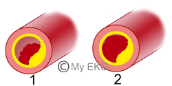
- 1. Partial occlusion of a coronary artery.
- 2. Complete occlusion of a coronary artery.
Subendocardial Injury Patern
Subendocardial injury is usually caused by a partial occlusion of a coronary artery, generating a greater degree of injury in subendocardium (more sensitive to ischemia) than subepicardium.
Subendocardial injury causes ST segment depression in more than one EKG lead. The ST depression is a sign of acute ischemia and appears in non-ST-elevation acute coronary syndrome (NSTE-ACS).
Subepicardial Injury Patern
When a total occlusion of a coronary artery occurs, a transmural injury appears (classically called subepicardial injury), This means that the entire myocardium in the area is affected.
The subepicardial injury produces ST segment elevation in leads near the affected regions 2 3.
Remember: ST elevation is a sign of acute myocardial infarction and must be treated as soon as possible (fibrinolysis or angioplasty). Read ST-segment elevation myocardial infarction...
Myocardial Infarction or Necrosis
Myocardial infarction or necrosis is caused by long-term persistence of ischemia. It usually appears in the evolution of ST-segment elevation myocardial infarction (STEMI), producing death (necrosis) of myocardial tissue.
The infarcted regions are electrically inactive, abnormal Q waves or QS complexes appear in leads near the affected regions.
Q wave infarction is usually transmural, because ischemic events limited to subendocardium do not cause Q wave (non Q wave infarction or NSTE-ACS).
Summary
Myocardial ischemia, injury and infarction are the different types of damage of myocardial tissues due to an acute coronary syndrome.
Each disturbance causes specific changes in the electrocardiogram, facilitating early diagnosis.
During the phases of ischemia and injury, myocardial injury is potentially reversible, but not at the phase of Infarction or necrosis.
We hope we have been of some help in expanding your knowledge. You may want to read this article acute myocardial nfarction.
References
- 1. Bayés de Luna A, Fiol-Sala M. La electrocardiografía de la cardiopatía isquémica. Correlaciones clínicas y de imagen e implicaciones pronósticas. Barcelona: Permanyer; 2012.
- 2. Wagner GS, Macfarlane P, Wellens H, et al. AHA/ACCF/HRS Recommendations for the Standardization and Interpretation of the Electrocardiogram: Part VI: Acute Ischemia/Infarction A Scientific Statement From the American Heart Association Electrocardiography and Arrhythmias Committee, Council on Clinical Cardiology; the American College of Cardiology Foundation; and the Heart Rhythm Society Endorsed by the International Society for Computerized Electrocardiology. J Am Coll Cardiol. 2009;53(11):1003-1011. doi:10.1016/j.jacc.2008.12.016
- 3. Rautaharju PM, Surawicz B, Gettes LS. AHA/ACCF/HRS Recommendations for the Standardization and Interpretation of the Electrocardiogram: Part IV: The ST Segment, T and U Waves, and the QT Interval A Scientific Statement From the American Heart Association Electrocardiography and Arrhythmias Committee, Council on Clinical Cardiology; the American College of Cardiology Foundation; and the Heart Rhythm Society Endorsed by the International Society for Computerized Electrocardiology. J Am Coll Cardiol. 2009;53(11):982-991. doi:10.1016/j.jacc.2008.12.014
If you Like it... Share it..


