Electrocardiogram Waves
Waves are the different upward or downward deflections represented on the EKG tracing. They are the product of the action potentials created during the cardiac stimulation, and repeated from one heart beat to another, barring alterations.
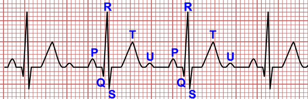
The electrocardiographic waves are called P, Q, R, S, T, U (in that order) and they are connected to each other by an isoelectric line.
P Wave
The P wave is the first wave of the cardiac cycle. It represents atrial depolarisation. It is the result of overlayiwg the electrical activity of both atria.
It wave duration is less that 0.10 s (2.5 mm wide) and its maximum amplitude is 0.25 mV (2.5 mm height). It is usually positive in all leads, except in aVR where it is negative, and in V1 where it is usually biphasic.
Atrial enlargements can bring about an increase in the P wave height or duration (read abnormal P wave). P wave is absent in atrial fibrillation.
Q Wave
Related article: The Q wave.
Two important things about this wave:
1. If there is a minimum positive wave in the QRS complex before a negative wave, the latter is not a Q wave but an S wave, no matter how small the previous positive wave.
2. Not every Q wave means infarction. On a normal electrocardiogram there are Q waves in certain leads without pathological significance.
Normal Q wave characteristics
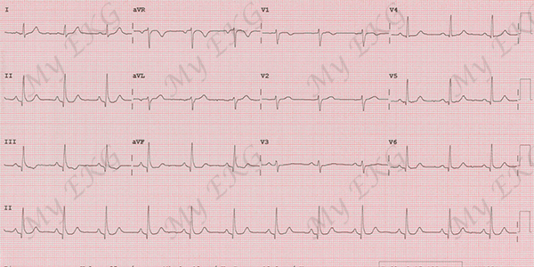
Normal Q wave:
Sinus rhythm at 72 bpm with non-pathologic Q wave in leads II, III, aVF, V4, V5 y V6.
Limb Leads:
- A normal Q wave is usually narrow and shallow (less that 0.04 s wide, 2 mm deep). Generally it does not exceed 25% of the QRS complex.
- A relatively deep Q wave can be seen in lead III in horizontally positioned hearts; a QS pattern can be seen in lead aVL in vertically positioned hearts.
- A deep Q wave is normal in lead aVF.
Precordial Leads:
- Q waves are not to be seen in leads V1-V2.
- Normally a Q wave can be seen in leads V5-V6, usually less that 0.04 s wide, 2 mm deep and it should never exceed 15% of the QRS complex.
More information: The Q wave.
QRS Complex
It consists of a collection of waves which represents the ventricular depolarisation. Its duration ranges from 0.06 s and 0.10 s. It can present different morphologies depending on the lead (read QRS complex morphology).
- Q wave: if the first wave of the QRS complex is negative, it is referred to as Q wave.
- R wave: it is the first positive wave in the QRS complex. It can be preceded by a negative wave. If another positive wave were present in the QRS complex, it is referred to as R’ wave.
- S wave: it is the second negative wave in the QRS complex, appearing after the R wave.
- QS wave: when a complex is completely negative, with absence of any positive wave, it is referred to as QS complex. It is usually a sign of necrosis.
- R' and S' waves: when more than a R or S wave are present, they are referred to as R’ wave and S’ wave.
Remember: if in the QRS complex there’s a minimal initial positive wave, no matter how small it might be, this is an R wave. The following negative wave should be an S wave, not a Q wave. Confusing the two is a common mistake.
T Wave
It represents ventricular repolarisation. In general it has a smaller amplitude than the QRS complex than precedes it.
It is positive in all leads, except in aVR. It could be negative in lead III in obese patients; in leads V1-V4 in children, young people and women.
The normal T wave is asymmetric, with an ascending portion which is slower than the descending one. Its maximum amplitud is less than 5 mm in limb leads and less than 15 mm in precordial leads.
There are many pathologies which cause changes in the T wave, such as coronary artery disease or electrolyte disorders (read abnormal T wave).
U Wave
It is a typically positive, low-voltage wave, which appears predominantly in precordial leads after the T wave. Its source is unknown, it could mean the repolarisation of the papillary muscles (read the U wave).
previous | next
If you Like it... Share it.