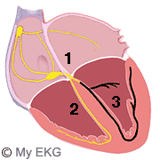
When the electrical stimulus from the AV node is not conducted by the left branch, a complete left bundle branch block (LBBB) occurs.
This interruption of the impulse must arise before the subdivison of the left branch, because the injury of a single fascicles causes a left fascicular block.
If both fascicles of the left branch are affected, left bundle branch block also occurs.

Left Bundle Branch Block
- 1. Atrioventricular node and bundle of His.
- 2. Normal right bundle branch.
- 3. Blocked left bundle branch.
In this condition, the electrical stimulation depolarize the left ventricle abnormally, because it is only conducted by the right branch.
The stimulus initially depolarized regions dependent on right branch (right ventricule and right third of the interventricular septum).
Then, the stimulus passes through the myocardium towards the left ventricle, increasing the time of ventricular depolarization (wide QRS complexes), resulting in abnormalities in the electrocardiogram.
Differences Between Normal Conduction and Left Bundle Branch Block
Normal conduction:
At the beginning of the QRS complex, a small R wave in lead V1, and a small Q wave in lead V6 are observed. They reflect the depolarization of the left septal region (first region to depolarize).
Subsequently, both ventricles are depolarized, with a predominance of the left ventricle, resulting in a deep S wave in lead V1 and a tall R wave in lead V6.
Left Bundle Branch Block:
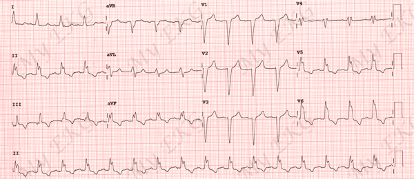
In left bundle branch block, the septum is depolarized from the right branch in right-left direction (contrary to normal), disappearing the initial R and Q waves in leads V1 and V6 respectively.
Subsequently, both ventricles are depolarized, with a predominance of left electrical forces, but total time for left ventricular depolarization is prolonged. This causes a negative and deep QRS comples (QS complex) in lead V1, and a wide and tall R wave in lead V6 2.
Differences between Normal EKG and Left Bundle Branch Block EKG:
Normal electrocardiogram: narrow QRS complex, lead V1 has an rS complex and lead V6 has a qR complex. T waves are normal.
Electrocardiogram with left bundle branch block: wide QRS, there is a broad QS complex in lead V1 and a broad notched or slurred R wave in leads V5 and V6.
In left bundle branch block there are also repolarization changes, secondary to abnormal depolarization. So, there is ST segment depression and negative T wave in precordial leads with tall R wave (V5 and V6).
Tip: T wave should be deflected opposite the terminal deflection of the QRS complex. Positive T wave in leads with upright QRS may be normal
Causes of Left Bundle Branch Block
Unlike right bundle branch block, left bundle branch block is usually associated with heart disease.
The main causes of left bundle branch block are:
- Left ventricular hypertrophy secondary to arterial hypertension or aortic stenosis.
- Ischemic heart disease.
- Valvular heart disease: aortic stenosis, mitral stenosis, aortic regurgitation.
- Dilated cardiomyopathy and hypertrophic cardiomyopathy.
- Conduction system disease (elderly patients).
- May appear at high heart rates (aberrant conduction).
- It may be observed in patients with completely normal cardiac studies.
Left bundle branch block may be the first evidence of undiagnosed heart disease. Every patient newly diagnosed with left bundle branch block requires complete cardiac evaluation.
Treatment of Left Bundle Branch Block
In the absence of underlying heart condition, left bundle branch block does not need treatment. If the LBBB was caused by a heart disease, this disease should be treated.
Patients with symptoms of acute coronary syndrome and unknown left bundle branch block must be treated as ST-segment elevation myocardial Infarction (primary angioplasty or fibrinolysis).
Patients with heart failure, left bundle branch block can cause left ventricular asynchrony (septum and lateral wall contract asynchronously), this condition often worsens the symptoms and the patient's prognosis. Resynchronization therapy usually improves ventricular function and reduces mortality of these patients.
Permanent pacemaker implantation is indicated for alternating bundle branch block (LBBB alternating with right bundle branch block)3.


